Correcting Eyelid Abnormalities: Surgical Techniques and Results
Some breeds wear their eye anatomy like a signature: the deep folds of the Shar-Pei, the dramatic drooping lower lids of the Basset Hound, the prominent eyes of the Bulldog. What reads as character in a photo can translate into chronic discomfort in real life, because eyelid conformation that causes the lid or lashes to contact the eye surface is not something a dog adapts to. It is something they live with, often squinting, tearing, and rubbing at their face, until someone decides to do something about it. Surgical correction of entropion and ectropion reliably resolves the problem when performed at the appropriate time and with proper technique.
Creature Comforts Veterinary Service in Saylorsburg, PA is an AAHA-accredited practice with 24/7 availability and comprehensive surgical capabilities that include the soft tissue procedures needed to correct eyelid abnormalities and restore comfortable, healthy eye function. Contact the team to evaluate a dog with chronic eye irritation that may have a structural cause.
When an Eye Problem Is Actually an Eyelid Problem
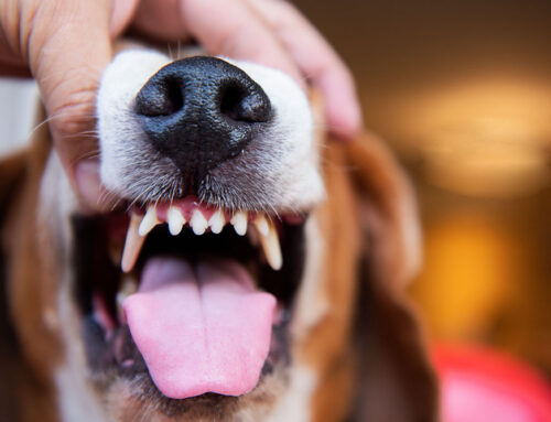
The signs are easy to notice and easy to dismiss: a dog who squints a little when you open the blinds, one eye that tears more than the other, a cat who occasionally rubs at their face. On their own, these might seem like minor things. In the context of eyelid disease, they’re the animal telling you their eye hurts.
Both entropion and ectropion are structural eyelid conditions that cause persistent eye irritation, and both are highly correctable with surgery. Understanding what each condition involves, which pets are at risk, and what treatment looks like helps owners make informed decisions when these problems arise rather than waiting until secondary damage has already occurred.
Entropion in dogs involves one or more eyelid margins rolling inward toward the eye. With every blink, the skin and lashes on the outer surface of the lid contact the cornea directly. The cornea is extremely sensitive tissue, and this constant contact is genuinely painful, producing tearing, squinting, and over time, potentially serious corneal damage.
Ectropion is the opposite condition: the lower eyelid droops outward and away from the eye, exposing the pink conjunctival tissue beneath. That exposed surface dries out, collects environmental debris, and becomes a reliable route for bacteria to trigger recurrent infections. The eye is chronically irritated even when nothing obviously wrong is visible from a distance.
Both conditions fall within the spectrum of common eye conditions that veterinary surgeons address regularly, but accurate diagnosis involves evaluating eyelid position, corneal surface health, tear production, and lash orientation together. Our team performs thorough ophthalmic evaluations and diagnostics before any surgical planning begins. Request an appointment today if your pet’s eyes have been bothering them, before it starts to impair their vision.
Which Breeds Are at Highest Risk?
Genetics and facial anatomy drive the majority of eyelid abnormality cases. Eyelid disorders occur at far higher rates in certain breed groups because their standard conformation includes the anatomical features that predispose to lid rolling or drooping. Hereditary eyelid conditions are well-documented and passed through breeding lines, which is part of why these problems concentrate so heavily in specific breeds.
Breeds most prone to entropion:
- Shar Peis, where extreme skin folds around the face create significant tension on the lids
- Chow Chows and Rottweilers
- English Bulldogs and Mastiffs
- Great Danes and Saint Bernards
- In cats, flat-faced breeds like Persians and Himalayans, whose compressed facial structure alters lid position
Breeds most prone to ectropion:
- Bloodhounds and Basset Hounds, where characteristically loose skin produces dramatic lower lid drooping
- Cocker Spaniels and Newfoundlands
- Saint Bernards and Great Danes
Some breeds, including Saint Bernards and Great Danes, can develop both conditions simultaneously in different portions of the same eyelid, a configuration sometimes referred to as “diamond eye” because of the shape the lid takes on. Contributing factors beyond breed include age-related tissue loosening in older pets, chronic eye inflammation that distorts lid position over time, previous injuries or surgeries, and squinting from pain that can temporarily pull a lid into an abnormal position.
Breed-specific eye concerns are worth discussing during wellness and preventive care visits for puppies and kittens in at-risk breeds, so owners know early what signs to monitor as the face matures.
Signs That Warrant a Veterinary Evaluation
An eye that looks even slightly off is worth getting checked, especially in a breed known for eyelid problems. Signs that suggest a structural eyelid issue include:
- Visible squinting or keeping one eye partially closed throughout the day
- Excessive tearing that stains the face or causes chronic wet fur below the eye
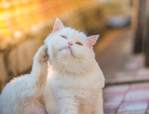
- Pawing or rubbing at the face, particularly the eye area
- Redness of the sclera (the white of the eye) or inner eyelid tissue
- Cloudy or hazy appearance of the normally clear cornea
- Crusting around the eye, mucus discharge, or recurrent eye infections
- A visible rolling of the lid edge inward or outward
These are all signs of eye pain that deserve prompt attention rather than watchful waiting. The common instinct to assume “that’s just how this breed looks” can delay treatment long enough for preventable corneal damage to occur. Request an evaluation if any of these signs are present.
What Happens When Eyelid Problems Go Untreated
The complications of untreated entropion and ectropion are progressive, and they get worse the longer they’re left.
For entropion, the ongoing friction of lash and lid tissue against the cornea causes corneal ulcers, which are open, painful sores on the eye surface. Superficial ulcers can be managed medically, but deep or infected ulcers may require surgical repair and carry a risk of perforation in severe cases. Chronic irritation also deposits pigment on the cornea over time, permanently reducing the transparency of the eye even after the lid problem is corrected.
For ectropion, the combination of exposed tissue, eye discharge, and chronic inflammation creates ideal conditions for bacterial infection. Repeated courses of antibiotics treat the infections temporarily, but the underlying structural problem keeps driving new ones. The longer these conditions continue without correction, the more complicated the surgical picture becomes and the less completely pre-existing damage resolves.
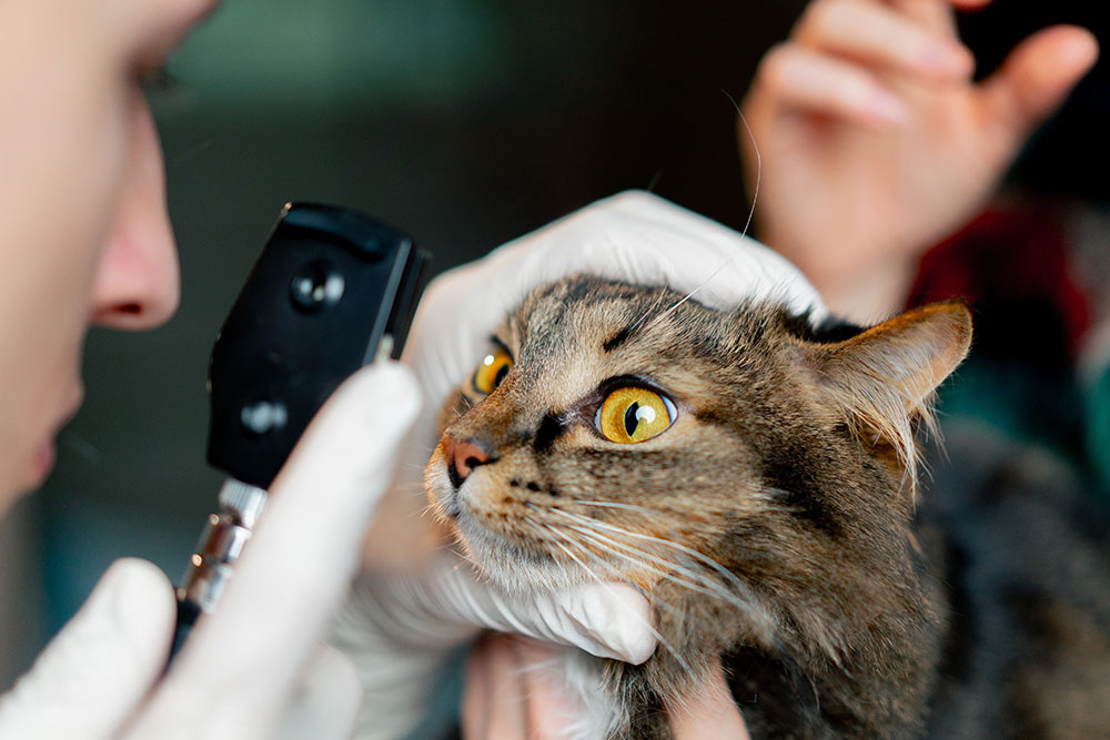
The Diagnostic Process: More Than Meets the Eye
Confirming an eyelid abnormality and planning surgical correction involves several specific steps that together build a complete picture of the eye and eyelid.
Applying topical anesthetic to the eye before examination is one of the most informative steps. Many dogs squint so intensely from eye pain that the squinting itself distorts the lid position. Numbing drops relax the squinting so the resting lid position can be evaluated accurately.
From there, a full ophthalmic workup includes:
- Tear production testing using a Schirmer tear test, which measures tear quantity using a small paper strip. Inadequate tear production can both mimic and worsen entropion symptoms.
- Fluorescein staining of the corneal surface to identify any existing ulcers or abrasions before surgery.
- Magnified lid and lash examination to check for eyelash problems like distichiasis or trichiasis, where abnormal lash placement produces entropion-like symptoms without actual lid rolling.
- Assessment of lid position and facial conformation to characterize the nature and degree of the abnormality.
Our diagnostics are conducted by a team experienced in identifying and differentiating these conditions accurately so that the surgical plan is based on what’s actually present.
Treatment: From Temporary Stabilization to Definitive Repair
When Surgery Can Wait
Not every dog with entropion needs immediate permanent correction. In puppies whose faces are still developing, the ideal lid position after the face reaches adult proportions can be hard to predict, which means correcting too early risks overcorrecting once growth is complete. For these dogs, temporary eyelid tacking is the appropriate first step: small sutures hold the eyelid in a more normal position, protecting the cornea from friction while the face matures. Tacking is a minor procedure performed under brief sedation or light anesthesia and can be repeated as needed. Our surgical team evaluates each patient carefully to determine whether immediate correction or a staged approach makes more sense.
Permanent Surgical Correction
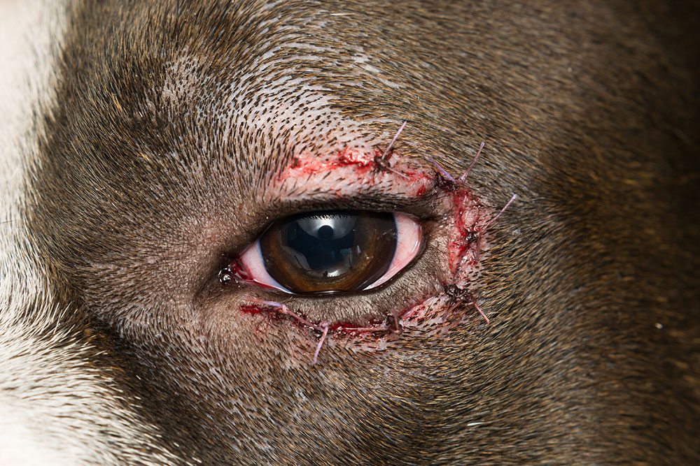
When the eyelid abnormality is clearly structural and the anatomy is stable, definitive surgery offers lasting relief. The specific technique depends on which condition is present, which portion of the lid is affected, how severe the abnormality is, and the individual patient’s anatomy. Both entropion and ectropion are corrected by precisely removing or repositioning a carefully measured amount of tissue to place the lid margin in normal contact with the eye.
The technical demands of eyelid surgery are significant because the margin for error is small. Too little tissue removed leaves the problem undercorrected; too much creates the opposite deformity. The preferred approach at Creature Comforts is conservative correction with the option to revisit if needed, rather than attempting aggressive correction in a single procedure. Anesthesia is fully individualized to each patient, with pre-anesthetic bloodwork, IV catheter placement for fluid support, and continuous monitoring of vitals throughout. As an AAHA-accredited practice, our anesthesia protocols meet rigorous independent standards.
Eyelid Conditions in Cats
Feline eyelid disease follows a somewhat different pattern than in dogs. Entropion in cats tends to develop later in life rather than being congenital, and it frequently occurs alongside chronic eye surface disease rather than as an isolated structural problem. Cats often require a combination of surgical approaches tailored specifically to their anatomy and to the concurrent eye conditions present. Cat owners wondering whether their pet’s eye problems might have a structural component are welcome to contact us directly.
What Surgery Day Looks Like at Creature Comforts
Walking through the day helps take some of the anxiety out of it.
- Bloodwork is reviewed before the procedure to confirm the pet is healthy for anesthesia, and the surgical plan is confirmed with any final questions answered.
- An individualized anesthesia protocol is prepared based on the pet’s age, breed, weight, and health status, with pain management incorporated before the procedure begins.
- The periocular area is prepared, precise measurements are taken, and the correction is executed with fine instruments and suture material, typically taking 30 to 60 minutes depending on complexity.
- A dedicated team member tracks heart rate, blood pressure, oxygen saturation, respiratory rate, and temperature continuously from induction through recovery.
- Most patients go home the same day once fully alert and stable, with written post-operative instructions and any needed medications.
Recovery at Home: The First Two Weeks
What’s Normal and What Warrants a Call
The first 24 to 48 hours bring the most visible changes. Mild swelling and bruising around the eyelid are expected and typically peak around the second day before beginning to resolve. A small amount of clear or slightly pink discharge from the eye is normal. Most pets are quieter than usual while pain medication is active.
Reach out to us if you notice:
- Swelling that is rapidly worsening rather than stable or improving
- Thick yellow or green discharge from the eye or incision
- Bleeding at the surgical site
- Sutures that are visibly loosened or wound edges that are separating
The E-collar is not negotiable during recovery. Eyelid sutures are small, precisely placed, and entirely at the mercy of one enthusiastic paw swipe. Tips on administering eye medications can make the post-operative drop and ointment routine much less stressful- having a second person hold the pet gently, following each application with a high-value treat, and keeping sessions brief helps most animals adjust within a few days. Contact us any time during recovery if something seems off.
The Healing Timeline
Recovery from eyelid surgery progresses in predictable stages:
- Days 10-14: Sutures are removed at a recheck appointment. The lid position at this point is close to the final result, though residual swelling may still be present.
- Weeks 3-6: All swelling resolves and the final corrected lid position becomes apparent. This is the clearest window for assessing the outcome.
- Follow-up as needed: Minor revisions are occasionally warranted after healing is complete, and the option to address them is straightforward when the original correction was conservative.
Post-operative rechecks are part of the process. Our wellness and preventive care team monitors ongoing eye health at annual exams for patients who have had eyelid corrections.
What Outcomes Look Like
Eyelid surgery has excellent outcomes when the patient is well-selected, the timing is right, and the home recovery is managed carefully. For most pets, the improvement in comfort is apparent quickly: the squinting diminishes, the tearing settles, and the constant face-rubbing stops.
Factors that influence the final result include the severity of lid malposition before surgery, the degree of corneal changes already present, the pet’s age and anatomy at the time of correction, and how consistently the cone and medications were used during recovery. Pre-existing corneal scarring or pigment deposits may not completely resolve after surgery, but eliminating the ongoing source of irritation prevents further damage and is a meaningful outcome in itself.
FAQ: Eyelid Surgery for Dogs and Cats
Can entropion come back after surgery?
Recurrence is uncommon when surgery is performed conservatively on an anatomically stable patient. In growing puppies treated with permanent correction too early, the continuing facial development can shift the lid position again, which is one reason timing matters so much. Staged correction in young dogs reduces this risk.
My cat has been squinting for months. Could it be entropion?
Possibly, though feline squinting has many causes including herpesvirus-related eye disease, which is extremely common in cats and can both cause and accompany entropion. A thorough ophthalmic evaluation distinguishes between structural and inflammatory causes, and many cats need treatment for both simultaneously.
Your Pet Deserves to See the World Comfortably
Eyelid abnormalities are among the most consistently correctable conditions in veterinary surgery, and the difference in a pet’s quality of life before and after correction is often dramatic. If your dog or cat is squinting, tearing chronically, or showing any of the signs discussed here, getting a proper evaluation is the right next step.
Creature Comforts Veterinary Service approaches every case, including eyelid surgery, with the same commitment: individualized medicine, thorough preparation, and a team genuinely invested in your pet’s outcome. We’re AAHA-accredited, available around the clock, and ready to help. Request an appointment online or call us at (570) 992-0400.

Leave A Comment