
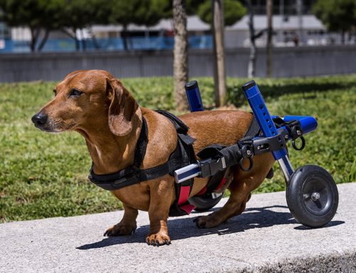
Degeneration and trauma to the back can lead to intervertebral disc disease (IVDD), which is the most common cause of paralysis in dogs, and can lead to permanent damage if not treated promptly. Our Creature Comforts Veterinary Service team provides information about this concerning condition to help you quickly recognize if your dog is affected and ensure treatment is not delayed.
Dog vertebrae anatomy
The dog’s backbone is composed of numerous small bones, called vertebrae, that form a protective, flexible casing for the spinal cord. Dogs have five cervical (i.e. neck) vertebrae, 13 thoracic (i.e. chest) vertebrae, seven lumbar (i.e. lower back) vertebrae, three fused sacral vertebrae, and a varying number of tail vertebrae. These bones are connected by intervertebral discs, which serve as cushioned joints between the vertebral bodies. Each intervertebral disc is composed of a hard outer shell (i.e., the annulus fibrosus) and a gel-like center (i.e., the nucleus pulposus). These discs, in conjunction with ligaments and muscles, help articulate the dog’s vertebral column to protect the spinal cord and spinal nerve roots.
Dog intervertebral disc disease types
Veterinarians have recognized IVDD in dogs since the 1800s, and research since that time has demonstrated two different disease processes that can lead to the condition:
- Hansen type I — Type I is most commonly seen in chondrodystrophoid breed dogs, such as dachshunds, Pekingese, beagles, and Lhasa apsos, and occurs when the nucleus pulposus becomes calcified, breaks through the annulus fibrosus, and puts pressure on the spinal cord. Signs typically present between 3 to 6 years of age.
- Hansen type II — Type II most commonly affects breeds such as German shepherds and Labrador retrievers, and is a much slower process that occurs when the annulus fibrosus degenerates and allows the nucleus pulposus to protrude and impinge on the spinal cord. Signs typically present between 5 to 12 years of age.
Most IVDD lesions occur in the thoracolumbar region, usually between thoracic vertebrae 11 and 12 and lumbar vertebrae two and three. Cervical lesions, which are most common in chondrodystrophoid breeds, typically occur between cervical vertebrae two and three.
Dog intervertebral disc disease signs
IVDD signs depend on the degree of disc herniation and the lesion location in the spinal column.
- Cervical lesions — IVDD in the cervical vertebrae causes signs including neck pain, reluctance to move the head and neck, lowered head stance, and weakness or partial paralysis in all four limbs. These lesions tend to create more pain but less function loss.
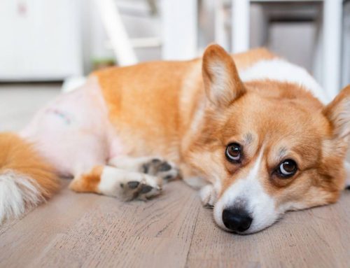
- Thoracolumbar lesions — IVDD in the thoracolumbar region can cause signs that vary greatly in severity. Mild signs include an unwillingness to jump or navigate stairs, reluctance to be handled, anxiety, and decreased activity level. More severe signs include lameness, a drunken gait, dragging the hind limbs, and loss of bladder or bowel control.
Dog intervertebral disc disease stages
IVDD in dogs is staged to help determine treatment approach and prognosis. Stages include:
- Stage 1 — Affected dogs experience pain in the lesion location, but have no neurological deficits.
- Stage 2 — Stage 2 IVDD causes mild neurological deficits, such as incoordination and knuckling of paws.
- Stage 3 — Affected dogs can move their limbs, but can’t walk or stand.
- Stage 4 — Stage 4 IVDD causes paralysis, but the dog can feel deep pain.
- Stage 5 — Affected dogs are paralyzed and can’t feel deep pain.
Dog intervertebral disc disease diagnosis
If your dog exhibits back pain or their mobility seems impaired, contact our Creature Comforts Veterinary Service team as soon as possible, because the sooner the problem is addressed, the better their prognosis. Tests to help diagnose the problem include:
- Neurological examination — Our veterinary team performs a complete physical exam, evaluating your pet’s gait, posture, and reflexes to help determine lesion location.
- Blood work — We may perform a complete blood count (CBC) and biochemistry profile to help determine your dog’s overall health status and rule out other potential causes.
- Plain X-rays — Our veterinary team performs plain X-rays to rule out fractures, dislocations, or other obvious bony abnormalities. When IVDD is present, plain X-rays can determine the lesion location in 50% to 75% of cases.
- Myelography — Advanced imaging involving a dye injected around the spinal cord may be necessary to properly visualize the lesion. In some cases, we may recommend a computed tomography (CT) scan or magnetic resonance imaging (MRI).
Dog intervertebral disc disease treatment

Treatment depends on the dog’s IVDD stage. Medical treatment is a reasonable option if your dog can walk and is not experiencing much pain, but surgery is typically recommended for dogs experiencing significant neurological deficits and extreme pain. Options include:
- Medical treatment — The most important aspect of IVDD medical management is confinement—severely restricting your dog’s activity is critical for spinal cord recovery. Large dogs can be kept in a small room with no furniture to prevent them from jumping, and small dogs should be kept in a crate or overturned playpen. Affected dogs must be strictly confined for at least three weeks, and possibly longer, depending on their condition. Other conservative treatment approaches include pain medication, icing the affected area, and physical therapy exercises.
- Surgical treatment — If your dog has significant pain or neurologic deficits, they will need surgery to remove pressure from the spinal cord. The surgical approach depends on lesion location.
IVDD is a serious condition and requires immediate veterinary attention. If you suspect your dog is experiencing back pain, or they are having difficulty moving, contact our Creature Comforts Veterinary Service team to ensure their condition is addressed promptly.

Leave A Comment